Bibliography

2017
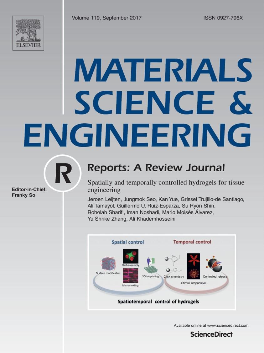
Spatially and temporally controlled hydrogels for tissue engineering.
Leijten J, Seo J, Yue K, Trujillo-de Santiago G, Tamayol A, Ruiz-Esparza GU, Shin SR, Sharifi R, Noshadi I, Álvarez MM, Zhang YS, and Khademhosseini A. 7/2017. “Spatially and temporally controlled hydrogels for tissue engineering.” Materials Science and Engineering: R: Reports. LinkAbstract
Recent years have seen tremendous advances in the field of hydrogel-based biomaterials. One of the most prominent revolutions in this field has been the integration of elements or techniques that enable spatial and temporal control over hydrogels’ properties and functions. Here, we critically review the emerging progress of spatiotemporal control over biomaterial properties towards the development of functional engineered tissue constructs. Specifically, we will highlight the main advances in the spatial control of biomaterials, such as surface modification, microfabrication, photo-patterning, and bioprinting, as well as advances in the temporal control of biomaterials, such as controlled release of molecules, photocleaving of proteins, and controlled hydrogel degradation. We believe that the development and integration of these techniques will drive the evolution of next-generation engineered tissues.
leijten_et_al_materials_science_and_engineering_r.pdf
A highly adhesive and naturally derived sealant.
Assmann A, Vegh A, Ghasemi-Rad M, Bagherifard S, Cheng G, Sani ES, Ruiz-Esparza GU, Noshadi I, Lassaletta AD, Gangadharan S, Tamayol A, Khademhosseini A, and Annabi N. 6/2017. “A highly adhesive and naturally derived sealant.” Biomaterials. LinkAbstract
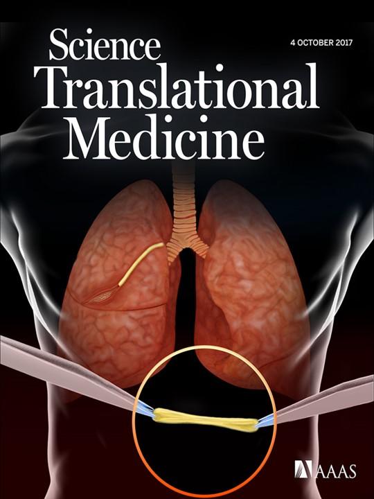
Conventional surgical techniques to seal and repair defects in highly stressed elastic tissues are insufficient. Therefore, this study aimed to engineer an inexpensive, highly adhesive, biocompatible, and biodegradable sealant based on a modified and naturally derived biopolymer, gelatin methacryloyl (GelMA). We tuned the degree of gelatin modification, prepolymer concentration, photoinitiator concentration, and crosslinking conditions to optimize the physical properties and adhesion of the photocrosslinked GelMA sealants. Following ASTM standard tests that target wound closure strength, shear resistance, and burst pressure, GelMA sealant was shown to exhibit adhesive properties that were superior to clinically used fibrin- and poly(ethylene glycol)-based glues. Chronic in vivo experiments in small as well as translational large animal models proved GelMA to effectively seal large lung leakages without the need for sutures or staples, presenting improved performance as compared to fibrin glue, poly(ethylene glycol) glue and sutures only. Furthermore, high biocompatibility of GelMA sealant was observed, as evidenced by a low inflammatory host response and fast in vivo degradation while allowing for adequate wound healing at the same time. Combining these results with the low costs, ease of synthesis and application of the material, GelMA sealant is envisioned to be commercialized not only as a sealant to stop air leakages, but also as a biocompatible and biodegradable hydrogel to support lung tissue regeneration.
assmann_et_al_biomaterials.pdf
Association of Irisin Plasma Levels with Anthropometric Parameters in Children with Underweight, Normal Weight, Overweight, and Obesity.
Elizondo-Montemayor L, Silva-Platas C, Torres-Quintanilla A, Rodriguez-Lopez C, Ruiz-Esparza GU, Reyes-Mendoza E, and Garcia-Rivas G. 5/3/2017. “Association of Irisin Plasma Levels with Anthropometric Parameters in Children with Underweight, Normal Weight, Overweight, and Obesity.” Biomed Research International. LinkAbstract

The correlations between irisin levels, physical activity, and anthropometric measurements have been extensively described in adults with considerable controversy, but little evidence about these relationships has been found in children. The objective of this study is to correlate the plasma levels of irisin in underweight, normal weight, overweight, and obese children with anthropometric parameters and physical activity levels. A cross-sample of 40 children was divided into the following groups on the basis of body mass index (BMI) percentile. The correlations of plasma irisin levels with physical activity, anthropometric, and metabolic measurements were determined. Plasma irisin levels (ng/mL) were lower for the underweight group (164.2 ± 5.95) than for the normal weight and obese groups (182.8 ± 5.58; ). Irisin levels correlated positively with BMI percentile (0.387), waist circumference (0.373), and fat-free mass (0.353; ), but not with body muscle mass (−0.027). After a multiple linear regression analysis, only BMI percentile (0.564; ) showed a positive correlation with irisin. Our results indicated no association with metabolic parameters. A negative correlation with physical activity was observed. Interrelationships among body components might influence irisin levels in children.

elizondo-montemayor_et_al_biomed_research_international.pdf
Paper-based microfluidic system for tear electrolyte analysis.
Yetisen AK, Jiang N, Tamayol A, Ruiz-Esparza GU, Zhang YS, Medina-Pando S, Gupta A, Wolffsohn JS, Butt H, Khademhosseini A, and Yun SH. 3/2017. “Paper-based microfluidic system for tear electrolyte analysis. .” Lab on a Chip. LinkAbstract
The analysis of tear constituents at point-of-care settings has a potential for early diagnosis of ocular disorders such as dry eye disease, low-cost screening, and surveillance of at-risk subjects. However, current minimally-invasive rapid tear analysis systems for point-of-care settings have been limited to assessment of osmolarity or inflammatory markers and cannot differentiate between dry eye subclassifications. Here, we demonstrate a portable microfluidic system that allows quantitative analysis of electrolytes in the tear fluid that is suited for point-of-care settings. The microfluidic system consists of a capillary tube for sample collection, a reservoir for sample dilution, and a paper-based microfluidic device for electrolyte analysis. The sensing regions are functionalized with fluorescent crown ethers, o-acetanisidide, and seminaphtorhodafluor that are sensitive to mono- and divalent electrolytes, and their fluorescence outputs are measured with a smartphone readout device. The measured sensitivity values of Na+, K+, Ca2+ ions and pH in artificial tear fluid were matched with the known ion concentrations within the physiological range. The microfluidic system was tested with samples having different ionic concentrations, demonstrating the feasibility for the detection of early-stage dry eye, differential diagnosis of dry eye sub-types, and their severity staging.
yetisen_et_al_lab_on_a_chip.pdf
Glucose-Sensitive Hydrogel Optical Fibers Functionalized with Phenylboronic Acid.
Yetisen AK, Jiang N, Fallahi A, Montelongo Y, Ruiz-Esparza GU, Tamayol A, Zhang YS, Mahmood I, Yang SA, Kim KS, Butt H, Khademhosseini A, and Yun SH. 2/2017. “Glucose-Sensitive Hydrogel Optical Fibers Functionalized with Phenylboronic Acid.” Advanced Materials. LinkAbstract

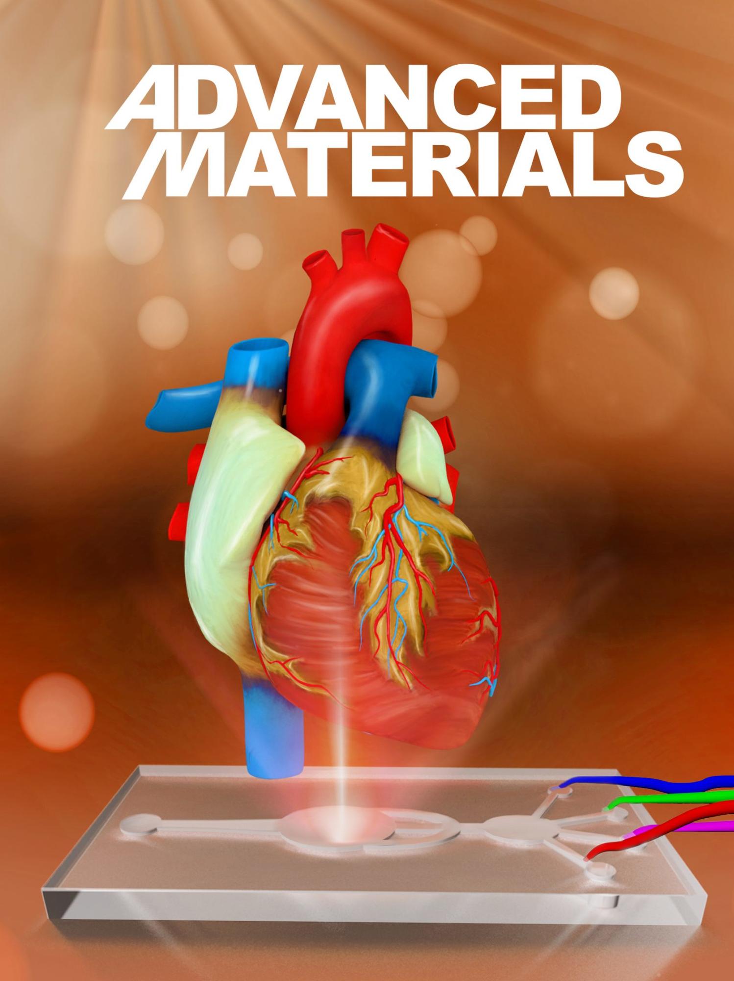
Hydrogel optical fibers are utilized for continuous glucose sensing in real time. The hydrogel fibers consist of poly(acrylamide-co-poly(ethylene glycol) diacrylate) cores functionalized with phenylboronic acid. The complexation of the phenylboronic acid and cis-diol groups of glucose enables reversible changes of the hydrogel fiber diameter. The analyses of light propagation loss allow for quantitative glucose measurements within the physiological range.

yetisen_et_al_advanced_materials.pdf
The commercialization of genome-editing technologies.
Brinegar K, Yetisen AK, Choi S, Vallillo E, Ruiz-Esparza GU, Prabhakar AM, Khademhosseini A, and Yun SH. 1/18/2017. “The commercialization of genome-editing technologies.” Critical Reviews in Biotechnology, Pp. 1-12. LinkAbstract
The emergence of new gene-editing technologies is profoundly transforming human therapeutics, agriculture, and industrial biotechnology. Advances in clustered regularly interspaced short palindromic repeats (CRISPR) have created a fertile environment for mass-scale manufacturing of cost-effective products ranging from basic research to translational medicine. In our analyses, we evaluated the patent landscape of gene-editing technologies and found that in comparison to earlier gene-editing techniques, CRISPR has gained significant traction and this has established dominance. Although most of the gene-editing technologies originated from the industry, CRISPR has been pioneered by academic research institutions. The spinout of CRISPR biotechnology companies from academic institutions demonstrates a shift in entrepreneurship strategies that were previously led by the industry. These academic institutions, and their subsequent companies, are competing to generate comprehensive intellectual property portfolios to rapidly commercialize CRISPR products. Our analysis shows that the emergence of CRISPR has resulted in a fivefold increase in genome-editing bioenterprise investment over the last year. This entrepreneurial movement has spurred a global biotechnology revolution in the realization of novel gene-editing technologies. This global shift in bioenterprise will continue to grow as the demand for personalized medicine, genetically modified crops and environmentally sustainable biofuels increases. However, the monopolization of intellectual property, negative public perception of genetic engineering and ambiguous regulatory policies may limit the growth of these market segments.
brinegar_et_al_critical_reviews_in_biotechnology.pdf
Development of an Elastic and Adhesive Sealant for Surgical Applications
N Annabi, Y. Zhang, A Assmann, Shirzaei E Sani, A Vegh, G. Cheng, B Dehghani, GU Ruiz-Esparza, X. Wang, AS Lassaletta, S Gangadharan, AS Weiss, and Khademhosseini A. 2017. “Development of an Elastic and Adhesive Sealant for Surgical Applications.” In Tissue Engineering Part A, 23: Pp. S123-S123. LinkAbstract
Conventional surgical sealants have been used for sealing or repairing defects often suffer from low adhesion strength, insufficient mechanical stability and strength, cytotoxic degradation products, and weak performance in biological environments. Therefore, in this study we aimed to engineer a photocrosslinked and highly biocompatible sealant with tunable mechanical and adhesion properties using tropoelastin, as a genetically modified human protein. We tuned the degree of methacrylation of tropoelastin and prepolymer concentration to optimize the physical properties and adhesion strength of the methacryloyl-substituted tropoelastin (MeTro) hydrogel for sealing of elastic and soft tissues. Following ASTM standard tests, the MeTro hydrogels revealed superior adhesive strength and burst pressure values compared to the commercially available sealants. The subcutaneous implantation of the engineered MeTro hydrogels in rats exhibited minimal inflammatory host responses and slow biodegradation of sealant. The in vivo and ex vivo burst pressure resistance of bioengineered MeTro sealants was tested on lungs and arteries in small as well as translational large animal models. Our results proved MeTro sealant to effectively seal lung and artery leakages without the need for sutures or staples, presenting a significant improvement compared to the commercially available clinical sealants (Evicel and ProgelTM) and sutures only. Combining these results, we envision that the engineered MeTro sealant has the potential to be commercialized due to its remarkable mechanical
strength, biocompatibility, biodegradability and strong adhesive interaction between the sealant and the wound tissue without the need for suturing.
annabi_et_al_tissue_engineering_part_a.pdf
2016
a
Ruiz-Esparza GU, Segura-Ibarra V, Cordero-Reyes AM, Youker KA, Serda RE, Cruz-Solbes AS, Amione-Guerra J, Yokoi K, Kirui DK, Cara FE, Paez-Mayorga J, Flores-Arredondo JH, Guerrero-Beltran CE, Garcia-Rivas G, Ferrari M, Blanco E, and Torre-Amione G. 1/2016. “A specifically designed nanoconstruct associates, internalizes, traffics in cardiovascular cells, and accumulates in failing myocardium: a new strategy for heart failure diagnostics and therapeutics.” European Journal of Heart Failure, 18, 2, Pp. 169-78. LinkAbstract

Aims

Ongoing inflammation and endothelial dysfunction occurs within the local microenvironment of heart failure, creating an appropriate scenario for successful use and delivery of nanovectors. This study sought to investigate whether cardiovascular cells associate, internalize, and traffic a nanoplatform called mesoporous silicon vector (MSV), and determine its intravenous accumulation in cardiac tissue in a murine model of heart failure.

Methods and results

In vitro cellular uptake and intracellular trafficking of MSVs was examined by scanning electron microscopy, confocal microscopy, time-lapse microscopy, and flow cytometry in cardiac myocytes, fibroblasts, smooth muscle cells, and endothelial cells. The MSVs were internalized within the first hours, and trafficked to perinuclear regions in all the cell lines. Cytotoxicity was investigated by annexin V and cell cycle assays. No significant evidence of toxicity was found. In vivo intravenous cardiac accumulation of MSVs was examined by high content fluorescence and confocal microscopy, with results showing increased accumulation of particles in failing hearts compared with normal hearts. Similar to observations in vitro, MSVs were able to associate, internalize, and traffic to the perinuclear region of cardiomyocytes in vivo.

Conclusions

Results show that MSVs associate, internalize, and traffic in cardiovascular cells without any significant toxicity. Furthermore, MSVs accumulate in failing myocardium after intravenous administration, reaching intracellular regions of the cardiomyocytes. These findings represent a novel avenue to develop nanotechnology-based therapeutics and diagnostics in heart failure.

ruiz-esparza_et_al_european_journal_of_heart_failure.pdf
2015
Nanotechnology-based delivery system for the transport of therapeutic and diagnostic molecules to the failing heart.
Ruiz Esparza GU, Cordero-Reyes A, KA Youker, Ferrari M, Blanco E, and Torre-Amione G. 5/2015. “Nanotechnology-based delivery system for the transport of therapeutic and diagnostic molecules to the failing heart.” Heart Failure 2015 / World Congress on Acute Heart Failure. Seville, Spain: European Society of Cardiology. LinkAbstract
Background: Treatment of heart failure has been limited by the inability to deliver high drug concentrations within the myocardium without significant systemic side effects and by the lack of efficient non-invasive methods for gene delivery into the myocardium. Nanotechnology platforms represent a potential strategy to transport drugs and genes in heart failure. We hypothesized that the changes present in the failing myocardium, result in increased vascular permeability due to endothelial dysfunction, allowing migration of intravenously administered silicon nanoconstructs from the vasculature to within the myocardium. 

Methods: Male C57BL/6 mice with normal and failing hearts were administered intravenously with silicon nanoconstructs (109, 0.2 mg).  After 24 h, mice were sacrificed and heart tissues were extracted and sectioned. To examine accumulation and homogeneity of the nanoconstructs in different cardiac regions, sections of normal and failing hearts were captured using wield field high content fluorescence imaging system. Cellular and sub-cellular localization of nanoconstructs was evaluated via immunofluorescence using confocal microscopy and z-stacking. Area fraction analysis and particle number was determined and quantified analyzing the fluorescence emitted by the nanoconstructs and the tissue. 

Results: Passive intra-cardiac accumulation of high concentrations of nanocarriers occurred after a single application during the course of 24 h.  Image analysis of the cardiac tissue showed intracellular uptake and transport of the nanoconstructs to the perinuclear region of cardiomyoctes. Control hearts showed slight green fluorescence associated with the nanoconstructs, whereas failing heart sections were highly enriched with fluorescence within different regions, indicating a 14-fold increase in accumulation of nanoconstructs. The highest levels of accumulation and co-localization of the nanoconstructs were present in cardiomyocytes. 

Conclusions: In summary, silicon nanoconstructs successfully accumulated in higher amounts in failing hearts compared to normal hearts, reaching the perinuclear region of the cardiomyocytes. The use of nanotechnology in heart failure, has the potential to achieve cardiac delivery of several moieties such as hydrophobic drugs, proteins, genetic material, or imaging and diagnostic agents in a safe, non-invasive, stable and protected fashion without degradation.
2014
Polymer Nanoparticles Encased in a Cyclodextrin Complex Shell for Potential Site- and Sequence-Specific Drug Release.
Ruiz-Esparza GU, Wu S, Segura-Ibarra V, Cara FE, Evans KW, Milosevic M, Ziemys A, Kojic M, Meric-Bernstam F, Ferrari M, and Blanco E. 8/13/2014. “Polymer Nanoparticles Encased in a Cyclodextrin Complex Shell for Potential Site- and Sequence-Specific Drug Release. .” Advanced Functional Materials, 24, 30, Pp. 4753-61. LinkAbstract

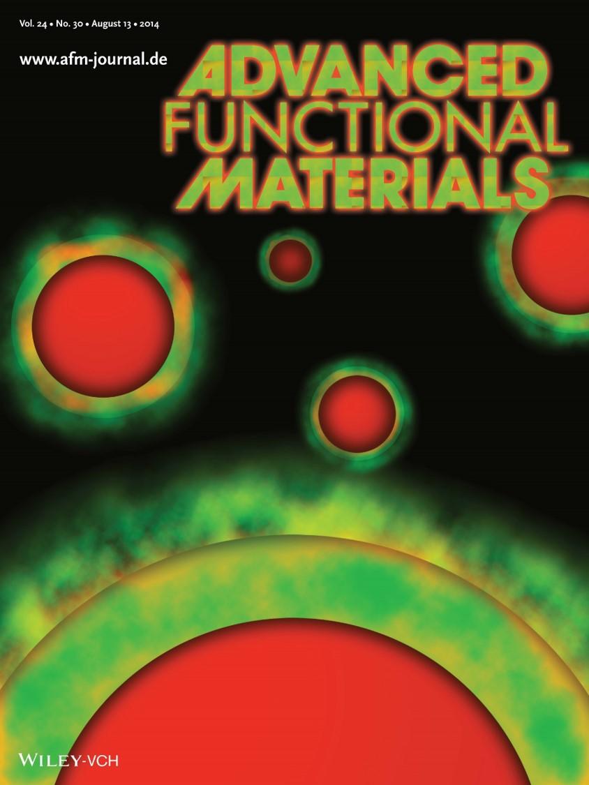
Time-staggered combination chemotherapy strategies show immense potential in cell culture systems, but fail to successfully translate clinically due to different routes of administration and disparate formulation parameters that preclude a specific order of drug presentation. A novel platform consisting of drug-containing PLGA polymer nanoparticles, stably fashioned with a shell composed of drug complexed with cationic cyclodextrin, capable of releasing drugs time- and sequence-specifically within tumors is designed. Morphological examination of nanoparticles measuring 150 nm highlight stable and distinct compartmentalization of model drugs, rhodamine and bodipy, within the core and shell, respectively. Sequential release is observed in vitro, owing to cyclodextrin shell displacement and subsequent sustained release of core-loaded drug, kinetics preserved in breast cancer cells following internalization. Importantly, time-staggered release is corroborated in a murine breast cancer model following intravenous administration. Precise control of drug release order, site-specifically, potentially opens novel avenues in polychemotherapy for synergy and chemosensitization strategies.

ruiz-esparza_et_al_advanced_functional_materials.pdf
Colocalized Delivery of Rapamycin and Paclitaxel to Tumors Enhances Synergistic Targeting of the PI3K/Akt/mTOR Pathway
Blanco E, Sangai T, Wu S, Hsiao A, Ruiz-Esparza GU, Gonzalez-Delgado CA, Cara FE, Granados-Principal S, Evans KW, Akcakanat A, Wang Y, Do KA, Meric-Bernstam F, and Ferrari M. 4/1/2014. “Colocalized Delivery of Rapamycin and Paclitaxel to Tumors Enhances Synergistic Targeting of the PI3K/Akt/mTOR Pathway.” Molecular Therapy, 22, 7, Pp. 1310-9. LinkAbstract

Ongoing clinical trials target the aberrant PI3K/Akt/mammalian target of rapamycin (mTOR) pathway in breast cancer through administration of rapamycin, an allosteric mTOR inhibitor, in combination with paclitaxel. However, synergy may not be fully exploited clinically because of distinct pharmacokinetic parameters of drugs. This study explores the synergistic potential of site-specific, colocalized delivery of rapamycin and paclitaxel through nanoparticle incorporation. Nanoparticle drug loading was accurately controlled, and synergistic drug ratios established in vitro. Precise drug ratios were maintained in tumors 48 hours after nanoparticle administration to mice, at levels twofold greater than liver and spleen, yielding superior antitumor activity compared to controls. Simultaneous and preferential in vivo delivery of rapamycin and paclitaxel to tumors yielded mechanistic insights into synergy involving suppression of feedback loop Akt phosphorylation and its downstream targets. Findings demonstrate that a same time, same place, and specific amount approach to combination chemotherapy by means of nanoparticle delivery has the potential to successfully translate in vitro synergistic findings in vivo. Predictive in vitro models can be used to determine optimum drug ratios for antitumor efficacy, while nanoparticle delivery of combination chemotherapies in preclinical animal models may lead to enhanced understanding of mechanisms of synergy, ultimately opening several avenues for personalized therapy.

blanco_et_al_molecular_therapy.pdf
2013
Cellular association of nanocarriers in cardiac cells.
Ruiz-Esparza GU, Blanco E, Shamsdueen S, Flores-Arredondo JH, Serda R, Ferrari M, and Torre-Amione G. 3/10/2013. “Cellular association of nanocarriers in cardiac cells.” In Journal of the American College of Cardiology, 10th ed., 61: Pp. E1834. San Francisco, California, USA: ACC.13 – American College of Cardiology 62nd Annual Scientific Session & Expo. LinkAbstract
Background: Mesoporous silicon particles (MSVs) offer delivery of nanotherapeutics in a sustained fashion, while biofunctionalization with targeting ligands enhances accumulation in target tissues. We hypothesize MSVs can be internalized by cardiac cells, after which they can deliver powerful payloads of therapeutics including siRNA and genes. Intracellular uptake of MSVs was investigated in cardiac myocytes (CM), cardiac fibroblasts (CF), cardiac smooth muscle cells (CSMC) and cardiac endothelial cells (CEC).
Methods: Negatively charged fluorescent nanoparticles (10nm) were loaded within pores of positively charged MSVs (1μmx400nm). In vitro uptake in a ratio of 25 MSVs per cell was evaluated after 3 hours of incubation via image stream analysis, as well as confocal and scanning electron microscopy.
Results: MSVs underwent cellular uptake, resulting in intracellular trafficking. Data obtained by image stream analysis allowed for quantification of MSV uptake rate as a function of cell area. The mean uptake count per cell was 18, 16 and 4 for CEC, CSMC and CF respectively. CM were subjectively quantified via microscopy resulting in a 25% enhancement of MSV capture compared with CSMC.
Conclusions: Cardiac cells can effectively internalize MSVs, with differences arising from cellular morphology and intracellular trafficking. Multifunctional delivery systems, molecularly targeted with high affinity ligands, could have vast potential in personalized delivery of therapeutics to cardiac cells.
ruiz-esparza_et_al_journal_of_the_american_college_of_cardiology.pdf
The physiology of cardiovascular disease and innovative liposomal platforms for therapy.
Ruiz-Esparza GU, Flores-Arredondo JH, Segura-Ibarra V, Torre-Amione G, Ferrari M, Blanco E, and Serda RE. 2013. “The physiology of cardiovascular disease and innovative liposomal platforms for therapy.” International Journal of Nanomedicine. , 8, Pp. 629-40. LinkAbstract
Heart disease remains the major cause of death in males and females, emphasizing the need for novel strategies to improve patient treatment and survival. A therapeutic approach, still in its infancy, is the development of site-specific drug-delivery systems. Nanoparticle-based delivery systems, such as liposomes, have evolved into robust platforms for site-specific delivery of therapeutics. In this review, the clinical impact of cardiovascular disease and the pathophysiology of different subsets of the disease are described. Potential pathological targets for therapy are introduced, and promising advances in nanotherapeutic cardiovascular applications involving liposomal platforms are presented.
ruiz-esparza_et_al_international_journal_of_nanomedicine.pdf
2012
Site-Specific, Concomitant Delivery of Rapamycin and Paclitaxel in Breast Cancer: Consequent Synergistic Efficacy Enhancement
Blanco E, Takafumi S, Hsiao A, Ruiz-Esparza GU, Ferrari M, and Meric-Bernstam F. 12/15/2012. “Site-Specific, Concomitant Delivery of Rapamycin and Paclitaxel in Breast Cancer: Consequent Synergistic Efficacy Enhancement.” In Cancer Research, 24th ed., 72: Pp. P6-11-03-P6-11-03. San Antonio, Texas, USA: Thirty-Fifth Annual CTRC‐AACR San Antonio Breast Cancer Symposium. LinkAbstract

Background: The phosphatidylinositol 3-kinase (PI3K)/Akt/mammalian target of rapamycin (mTOR) (PI3k/Akt/mTOR) pathway is dysregulated in certain breast cancers. Ongoing clinical trials aim to therapeutically exploit this pathway through administration of rapamycin (RAP), an mTOR inhibitor, in combination with paclitaxel (PTX). However, actual drug synergy in clinical settings may not be fully realized due to disparate pharmacokinetic parameters of individual drug formulations, wherein drugs or their effects may never be present in the tumor at the same time. Our objective was to generate a nanoparticle platform capable of site-specifically delivering precise amounts of rapamycin and paclitaxel to breast tumors with hopes of increasing synergistic targeting of the PI3k/Akt/mTOR pathway.

Materials and Methods: Drug-containing nanoparticles composed of amphiphilic block copolymers of pegylated poly(∈-caprolactone) (PEG-PCL, MW = 5k-5k) were fabricated and nanoparticle size and drug loading efficiency was determined. In vitro growth inhibition of nanoparticle formulations of varying ratios was evaluated in MCF-7 and MDA-MB-468 breast cancer cells via sulforhodamine B assays, after which median-effect plot analyses and combination index calculations were conducted. Antitumor efficacy studies were performed in female nude mice bearing MDA-MB-468 tumors, in which nanoparticles were administered intravenously twice a week for the duration of three weeks. Biodistribution of drug-containing nanoparticles in extracted tumors were examined, as well as reverse phase protein array (RPPA) analysis to gain insights into site-specific synergy.

Results: Nanoparticles were spherical, with an average diameter of 9 nm. Both rapamycin and paclitaxel loaded favorably, allowing for customization of different ratios within nanoparticles. Combination indices demonstrated that a 3:1 ratio of RAP:PTX had the most synergy in MDA-MB-468 breast cancer cells in vitro, a synergy found to be preserved in vivo. Significant tumor regression (> 1.5 fold reduction from initial tumor volume) was observed in vivo upon administration of 3:1 RAP:PTX (15:5 mg/kg) nanoparticles. The precise ratio of rapamycin and paclitaxel (3:1) was found maintained in tumors 24 h after administration, an effect not achievable with free drug formulations. RPPA analysis demonstrated effective blocking of mTOR and Akt 24 h after administration of nanoparticles, key events in drug synergy.

Discussion: Site-specific delivery of synergistic agents in precisely-controlled drug ratios, possible through their incorporation into nanoparticles, was shown to be highly efficacious against breast tumors. Findings demonstrate the ability to deliver specific drug ratios to tumors, potentially precluding the need to administer maximal doses of both agents in order to achieve synergy, lessening patient side-effects. This study demonstrates the potential for prediction of in vivo therapeutic outcomes from in vitro synergistic findings. Nanoparticle delivery of drugs may also yield enhanced understanding of mechanisms of synergy between molecular-targeted drugs and traditional chemotherapeutics in vivo, resulting in novel and more efficacious treatment regimens.

blanco_et_al_cancer_research.pdf
2011
Mesoporous Silicon-Based Antigen Delivery Constructs for Cancer Vaccine Prevention and Therapy.
Loo CH, Meraz IM, Anderson HA, Qin G, DeLaCerda J, Ruiz-Esparza GU, Rubio-Calva V, Diaz LA, Li KC, and Serda RE. 11/2011. “Mesoporous Silicon-Based Antigen Delivery Constructs for Cancer Vaccine Prevention and Therapy.” In BMES Annual Meeting 11/2011. Hartford, Connecticut, USA: Biomedical Engineering Society.
Molecular-targeted nanotherapies in cancer: enabling treatment specificity.
Blanco E, Hsiao A, Ruiz-Esparza GU, Landry MG, Meric-Bernstam F, and Ferrari M. 10/13/2011. “Molecular-targeted nanotherapies in cancer: enabling treatment specificity.” Molecular Oncology, 5, 6, Pp. 492-503. LinkAbstract
Chemotherapy represents a mainstay and powerful adjuvant therapy in the treatment of cancer. The field has evolved from drugs possessing all-encompassing cell-killing effects to those with highly targeted, specific mechanisms of action; a direct byproduct of enhanced understanding of tumorigenic processes. However, advances regarding development of agents that target key molecules and dysregulated pathways have had only modest impacts on patient survival. Several biological barriers preclude adequate delivery of drugs to tumors, and remain a formidable challenge to overcome in chemotherapy. Currently, the field of nanomedicine is enabling the delivery of chemotherapeutics, including repositioned drugs and siRNAs, by giving rise to carriers that provide for protection from degradation, prolonged circulation times, and increased tumor accumulation, all the while resulting in reduced patient morbidity. This review aims to highlight several innovative, nanoparticle-based platforms with the potential of providing clinical translation of several novel chemotherapeutic agents. We will also summarize work regarding the development of a multistage drug delivery strategy, a robust carrier platform designed to overcome several biological barriers while en route to tumors.
Blanco_et_al_molecular_oncololy.pdf

Pages