Bibliography

2022
Polymers for surgical sutures
Afewerki S, Vargas Harb S, Domingues Stocco T, Ruiz-Esparza GU, and Lobo AO. 10/2/2022. “Polymers for surgical sutures.” In Advanced Technologies and Polymer Materials for Surgical Sutures. Cambridge: Elsevier. LinkAbstract
One of the biggest challenges within the medical practice is the innovation and improvements in technologies for the closure of wounds or sutures. The general technologies comprise physically perforating materials, for example, staples or sutures. These approaches have several limitations and challenges such as the risk of infections, cause continues pain, not always effective and in some cases can result in leakage at the site of closure. To overcome these challenges and limitations polymer-based sutures can be employed to hold body tissues together or ligate blood vessels, after a surgery or accidental injury. Depending on the damaged site, specific features are required to withstand the natural conditions of the body, but the utmost property for a suture material is its tensile strength, which can be tailored by the composition and thickness of the yarn. Aside the strength, other important properties to be considered are absorbability, sterility, high knot security, lack of allergic reaction, and ease of handling. In addition to these characteristics of biomaterials, in general, other criteria used for suture selection are based on the properties of the tissues involved, such as the specific healing rate; wound condition and general health of the patient, potential postoperative complications, personal preference and experience of the surgeon, and economic reasons. Among the extensive portfolio of materials currently available, synthetic and natural polymers have been the most frequently targeted.
afewerki_et_al_polymers_for_surgical_sutures.pdf
In vitro high-content tissue models to address precision medicine challenges
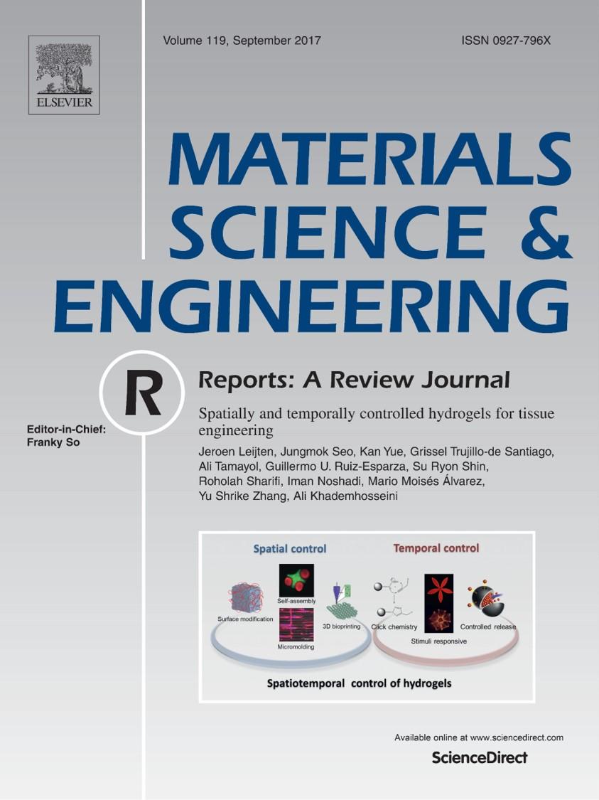
Afewerki S, Domingues Stocco T, Diniz Rosa Silva da A, Sales Aguiar Furtado A, Fernandes Sousa de G, Ruiz-Esparza GU, Webstere TJ, Marciano FR, Strømme M, Zhang YS, and Lobo AO. 8/1/2022. “In vitro high-content tissue models to address precision medicine challenges.” Molecular Aspects of Medicine. LinkAbstract
The field of precision medicine allows for tailor-made treatments specific to a patient and thereby improve the efficiency and accuracy of disease prevention, diagnosis, and treatment and at the same time would reduce the cost, redundant treatment, and side effects of current treatments. Here, the combination of organ-on-a-chip and bioprinting into engineering high-content in vitro tissue models is envisioned to address some precision medicine challenges. This strategy could be employed to tackle the current coronavirus disease 2019 (COVID-19), which has made a significant impact and paradigm shift in our society. Nevertheless, despite that vaccines against COVID-19 have been successfully developed and vaccination programs are already being deployed worldwide, it will likely require some time before it is available to everyone. Furthermore, there are still some uncertainties and lack of a full understanding of the virus as demonstrated in the high number new mutations arising worldwide and reinfections of already vaccinated individuals. To this end, efficient diagnostic tools and treat- ments are still urgently needed. In this context, the convergence of bioprinting and organ-on-a-chip technologies, either used alone or in combination, could possibly function as a prominent tool in addressing the current pandemic. This could enable facile advances of important tools, diagnostics, and better physiologically repre- sentative in vitro models specific to individuals allowing for faster and more accurate screening of therapeutics evaluating their efficacy and toxicity. This review will cover such technological advances and highlight what is needed for the field to mature for tackling the various needs for current and future pandemics as well as their relevancy towards precision medicine.
afewerki_et_al_molecular_aspects_of_medicine.pdf
From Bench to the Clinic: The Path to Translation of Nanotechnology-Enabled mRNA SARS-CoV-2 Vaccines
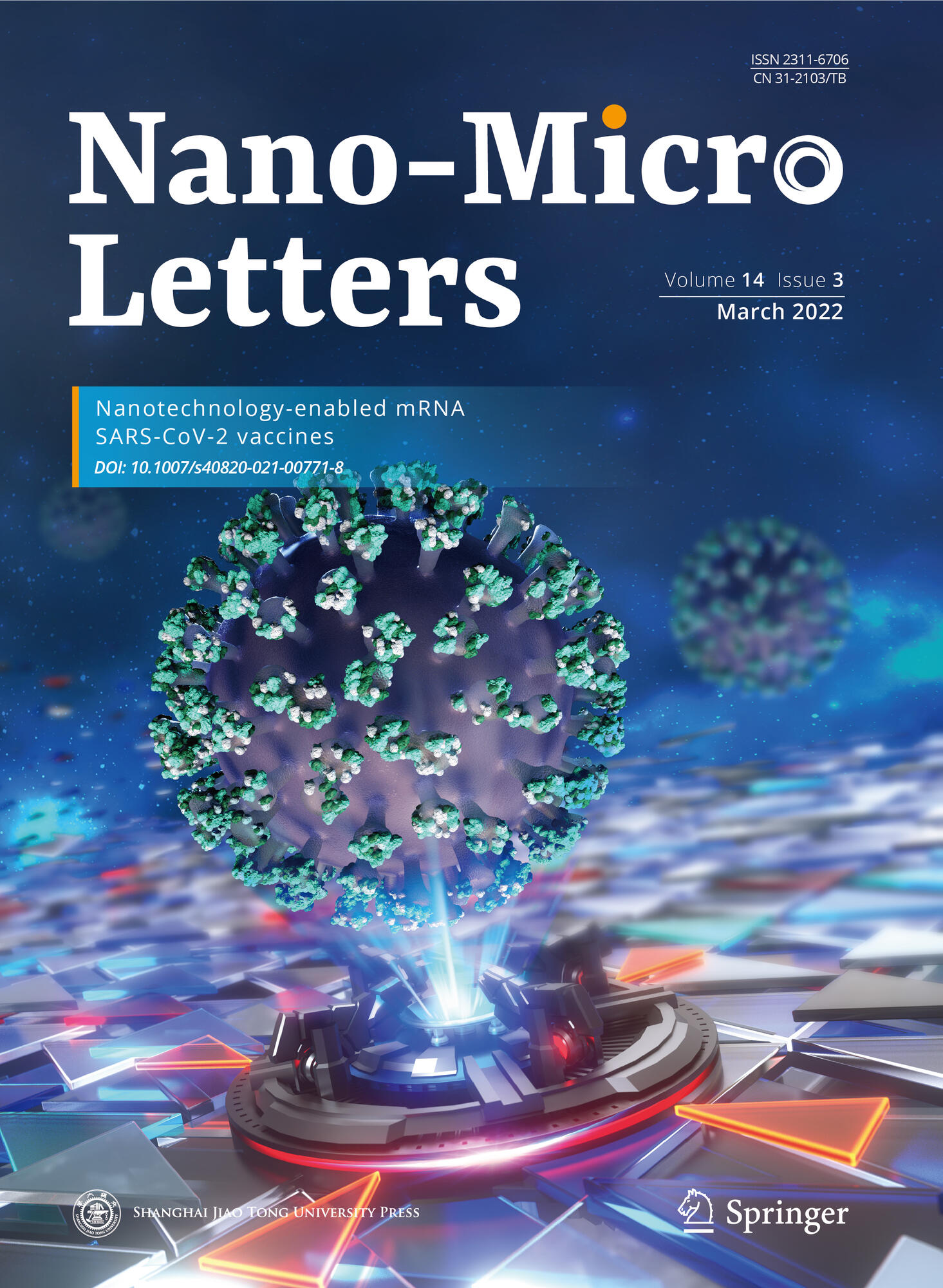
Lopez‐Cantu DO, Wang X, Carrasco‐Magallanes H, Afewerki S, Zhang X, Bonventre JV, and Ruiz-Esparza GU. 1/3/2022. “From Bench to the Clinic: The Path to Translation of Nanotechnology-Enabled mRNA SARS-CoV-2 Vaccines.” Nano-Micro Letters. LinkAbstract
During the last decades, the use of nanotechnology in medicine has effectively been translated to the design of drug delivery systems, nanostructured tissues, diagnostic platforms, and novel nanomaterials against several human diseases and infectious pathogens. Nanotechnology-enabled vaccines have been positioned as solutions to mitigate the pandemic outbreak caused by the novel pathogen severe acute respiratory syndrome coronavirus 2. To fast-track the development of vaccines, unprecedented industrial and academic collaborations emerged around the world, resulting in the clinical translation of effective vaccines in less than one year. In this article, we provide an overview of the path to translation from the bench to the clinic of nanotechnology-enabled messenger ribonucleic acid vaccines and examine in detail the types of delivery systems used, their mechanisms of action, obtained results during each phase of their clinical development and their regulatory approval process. We also analyze how nanotechnology is impacting global health and economy during the COVID-19 pandemic and beyond.
lopez-cantu_nano_micro_letters.pdf
2021
A Tissue-Bioengineering Strategy for Modeling Rare Human Kidney Diseases In Vivo
Hernandez J, Wang X, Vazquez-Segoviano M, Sobral-Reyes MF, Moran-Horowich A, Sundberg M, Lopez-Marfil M, Lopez-Cantu DO, Probst C, Ruiz-Esparza GU, Giannikou K, Henske E, Kwiatkowski D, Sahin M, and Lemos DR. 11/11/2021. “A Tissue-Bioengineering Strategy for Modeling Rare Human Kidney Diseases In Vivo.” Nature Communications. LinkAbstract
The lack of animal models for some human diseases precludes our understanding of disease mechanisms and our ability to test prospective therapies in vivo. Generation of kidney organoids from Tuberous Sclerosis Complex (TSC) patient-derived-hiPSCs allows us to recapitulate a rare kidney tumor called angiomyolipoma (AML). Organoids derived from TSC2−/− hiPSCs but not from isogenic TSC2+/− or TSC2+/+ hiPSCs share a common transcriptional signature and a myomelanocytic cell phenotype with kidney AMLs, and develop epithelial cysts, replicating two major TSC-associated kidney lesions driven by genetic mechanisms that cannot be consistently recapitulated with transgenic mice. Transplantation of multiple TSC2−/− renal organoids into the kidneys of immunodeficient rats allows us to model AML in vivo for the study of tumor mechanisms, and to test the efficacy of rapamycin-loaded nanoparticles as an approach to rapidly ablate AMLs. Collectively, our experimental approaches represent an innovative and scalable tissue-bioengineering strategy for modeling rare kidney disease in vivo.
hernandez_et_al_nature_communications.pdf
Nanoengineered Shear-thinning Hydrogel Barrier for Preventing Postoperative Adhesions
Ruiz-Esparza GU, Wang X, Zhang X, Jimenez-Vazquez S, Diaz-Gomez L, Lavoie AM, Afewerki S, Fuentes A, Parra-Saldivar R, Jiang N, Annabi N, Saleh B, Yetisen AK, Sheikhi A, Jozefiak TH, Shin SR, Dong N, and Khademhosseini A. 10/18/2021. “Nanoengineered Shear-thinning Hydrogel Barrier for Preventing Postoperative Adhesions.” Nano-Micro Letters. LinkAbstract
More than 90% of surgical patients develop postoperative adhesions, and the incidence of hospital re-admissions can be as high as 20%. Current adhesion barriers present limited efficacy due to difficulties in application and incompatibility with minimally invasive interventions. To solve this clinical limitation, we developed an injectable and sprayable shear-thinning hydrogel barrier (STHB) composed of silicate nanoplatelets and poly(ethylene oxide). We optimized this technology to recover mechanical integrity after stress, enabling its delivery though injectable and sprayable methods. We also demonstrated limited cell adhesion and cytotoxicity to STHB compositions in vitro. The STHB was then tested in a rodent model of peritoneal injury to determine its efficacy preventing the formation of postoperative adhesions. After two weeks, the peritoneal adhesion index was used as a scoring method to determine the formation of postoperative adhesions, and STHB formulations presented superior efficacy compared to a commercially available adhesion barrier. Histological and immunohistochemical examination showed reduced adhesion formation and minimal immune infiltration in STHB formulations. Our technology demonstrated increased efficacy, ease of use in complex anatomies, and compatibility with different delivery methods, providing a robust universal platform to prevent postoperative adhesions in a wide range of surgical interventions.
ruiz-esparza_et_al_nano_micro_letters.pdf
Molecularly Imprinted Polymer Nanogels for Protein Recognition: Direct Proof of Specific Binding Sites by Solution STD and WaterLOGSY NMR Spectroscopies
Mier A, Maffucci I, Merlier F, Prost E, Montagna E, Ruiz-Esparza GU, Bonventre JV, Dhal PK, Tse Sum Bui B, Sakhaii P, and Haupt K. 7/22/2021. “Molecularly Imprinted Polymer Nanogels for Protein Recognition: Direct Proof of Specific Binding Sites by Solution STD and WaterLOGSY NMR Spectroscopies.” Angewandte Chemie International Edition. LinkAbstract
Molecularly imprinted polymers (MIPs) are tailor-made synthetic antibodies possessing specific binding cavities designed for a target molecule. Currently, MIPs for protein targets are synthesized by imprinting a short surface-exposed fragment of the protein, called epitope or antigenic determinant. However, finding the epitope par excellence that will yield a peptide ‘synthetic antibody’ cross-reacting exclusively with the protein from which it is derived, is not easy. We propose a computer-based rational approach to unambiguously identify the ‘best’ epitope candidate. Then, using Saturation Transfer Difference (STD) and WaterLOGSY NMR spectroscopies, we prove the existence of specific binding sites created by the imprinting of this peptide epitope in the MIP nanogel. The optimized MIP nanogel could bind the epitope and cognate protein with a high affinity and selectivity. The study was performed on Hepatitis A Virus Cell Receptor-1 protein, also known as KIM-1 and TIM-1, for its ubiquitous implication in numerous pathologies.
mier_et_al_angewandte_chemie.pdf
Nanostructured Non-Newtonian Drug Delivery Barrier Prevents Postoperative Intrapericardial Adhesions
Wang X, Liu Z, Sandoval-Salaiza D, Afewerki S, Jimenez-Rodriguez MG, Sanchez-Melgar L, Güemes-Aguilar G, Gonzalez-Sanchez DG, Noble O, Lerma C, Parra-Saldivar R, Lemos DR, Llamas-Esperon GA, Shi J, Li L, Lobo AO, Fuentes-Baldemar AA, Bonventre JV, Dong N, and Ruiz-Esparza GU. 6/17/2021. “Nanostructured Non-Newtonian Drug Delivery Barrier Prevents Postoperative Intrapericardial Adhesions.” ACS Applied Materials & Interfaces. LinkAbstract
With the increasing volume of cardiovascular surgeries and the rising adoption rate of new methodologies that serve as a bridge to cardiac transplantation and that require multiple surgical interventions, the formation of postoperative intrapericardial adhesions has become a challenging problem that limits future surgical procedures, causes serious complications, and increases medical costs. To prevent this pathology, we developed a nanotechnology-based self-healing drug delivery hydrogel barrier composed of silicate nanodisks and polyethylene glycol with the ability to coat the epicardial surface of the heart without friction and locally deliver dexamethasone, an anti-inflammatory drug. After the fabrication of the hydrogel, mechanical characterization and responses to shear, strain, and recovery were analyzed, confirming its shear-thinning and self-healing properties. This behavior allowed its facile injection (5.75 ± 0.15 to 22.01 ± 0.95 N) and subsequent mechanical recovery. The encapsulation of dexamethasone within the hydrogel system was confirmed by 1H NMR, and controlled release for 5 days was observed. In vitro, limited cellular adhesion to the hydrogel surface was achieved, and its anti-inflammatory properties were confirmed, as downregulation of ICAM-1 and VCAM-1 was observed in TNF-α activated endothelial cells. In vivo, 1 week after administration of the hydrogel to a rabbit model of intrapericardial injury, superior efficacy was observed when compared to a commercial adhesion barrier, as histological and immunohistochemical examination revealed reduced adhesion formation and minimal immune infiltration of CD3+ lymphocytes and CD68+ macrophages, as well as NF-κβ downregulation. We presented a novel nanostructured drug delivery hydrogel system with unique mechanical and biological properties that act synergistically to prevent cellular infiltration while providing local immunomodulation to protect the intrapericardial space after a surgical intervention.
wang_et_al_acs_applied_materials_and_interfaces.pdf
A Nanostructured Non-Newtonian Drug Delivery Barrier Prevents Postoperative Intrapericardial Adhesions
Wang X and Ruiz-Esparza GU. 5/17/2021. “A Nanostructured Non-Newtonian Drug Delivery Barrier Prevents Postoperative Intrapericardial Adhesions.” Journal of the American College of Cardiology 77 (18), Pp. (Supplement_1) 3269. LinkAbstract

Background: With the increasing volume of cardiovascular surgeries and the adoption of new methodologies as a bridge to cardiac transplantation, the formation of postoperative intrapericardial adhesions limits future surgical procedures, causes serious complications, and increases medical costs. Currently, no technology specifically designed to prevent intrapericardial adhesions exists.

Methods: Rheological analysis of hydrogel compositions was performed by an AR-G2 rheometer. The injection force was quantified by a mechanical tester. The 1H-Nuclear Magnetic Resonance (NMR) analysis was tested to confirm the hydrogel components. The cytotoxicity and cell-material interaction of hydrogel compositions were evaluated with NIH 3T3 fibroblasts and mouse brain endothelial cells. Rabbit intrapericardial adhesions model was used to test the therapeutic efficacy of the hydrogel formulations in vivo.

Results: Mechanical characterization and hydrogel response to shear, strain, and recovery confirmed its shear-thinning and self-healing properties. This behavior allowed its facile injection and subsequent mechanical recovery. Dexamethasone hydrogel encapsulation and controlled release for 5 days was achieved. In vitro, biocompatibility and limited fibroblast adhesion to the hydrogel surface was observed, and its anti-inflammatory properties were confirmed. In vivo, after one week of administration to a rabbit model of intrapericardial injury, the hydrogel showed superior efficacy compared to a commercial adhesion barrier, as histological and immunohistochemical examination revealed reduced adhesion formation and minimal immune response. The echocardiographic assessment showed normal cardiac function after hydrogel administration.

Conclusion: We present a novel nanostructured hydrogel system with unique mechanical and biological properties that act synergistically to prevent cellular infiltration while providing local immunomodulation to protect the intrapericardial space after surgical intervention. This technology significantly reduces adhesion formation, resulting in a promising approach that could improve surgical cardiovascular outcomes.

wang_et_al_journal_of_the_american_college_of_cardiology.pdf
Engineering multifunctional bactericidal nanofibers for abdominal hernia repair
Afewerki S, Bassous N, Vargas Harb S, Corat MAF, Maharjan S, Ruiz-Esparza GU, Paula de MMM, Webster TJ, Tim CR, Cruz Viana B, Wang D, Wang X, Marciano FR, and Lobo AO. 2/19/2021. “Engineering multifunctional bactericidal nanofibers for abdominal hernia repair.” Communications Biology, 4. LinkAbstract
The engineering of multifunctional surgical bactericidal nanofibers with inherent suitable mechanical and biological properties, through facile and cheap fabrication technology, is a great challenge. Moreover, hernia, which is when organ is pushed through an opening in the muscle or adjacent tissue due to damage of tissue structure or function, is a dire clinical challenge that currently needs surgery for recovery. Nevertheless, post-surgical hernia complications, like infection, fibrosis, tissue adhesions, scaffold rejection, inflammation, and recurrence still remain important clinical problems. Herein, through an integrated electrospinning, plasma treatment and direct surface modification strategy, multifunctional bactericidal nanofibers were engineered showing optimal properties for hernia repair. The nanofibers displayed good bactericidal activity, low inflammatory response, good biodegradation, as well as optimal collagen-, stress fiber- and blood vessel formation and associated tissue ingrowth in vivo. The disclosed engineering strategy serves as a prominent platform for the design of other multifunctional materials for various biomedical challenges.
afewerki_et_al_communications_biology.pdf
2020
Combined Catalysis for Engineering Bioinspired, Lignin-Based, Long-Lasting, Adhesive, Self-Mending, Antimicrobial Hydrogels
Afewerki S, Wang X, Ruiz-Esparza GU, Tai C, Kong X, Zhou S, Welch K, Huang P, Bengtsson R, Xu C, and Strømme M. 12/11/2020. “Combined Catalysis for Engineering Bioinspired, Lignin-Based, Long-Lasting, Adhesive, Self-Mending, Antimicrobial Hydrogels.” ACS Nano. LinkAbstract
The engineering of multifunctional biomaterials using a facile sustainable methodology that follows the principles of green chemistry is still largely unexplored but would be very beneficial to the world. Here, the employment of catalytic reactions in combination with biomass-derived starting materials in the design of biomaterials would promote the development of eco-friendly technologies and sustainable materials. Herein, we disclose the combination of two catalytic cycles (combined catalysis) comprising oxidative decarboxylation and quinone-catechol redox catalysis for engineering lignin-based multifunctional antimicrobial hydrogels. The bioinspired design mimics the catechol chemistry employed by marine mussels in nature. The resultant multifunctional sustainable hydrogels (1) are robust and elastic, (2) have strong antimicrobial activity, (3) are adhesive to skin tissue and various other surfaces, and (4) are able to self-mend. A systematic characterization was carried out to fully elucidate and understand the facile and efficient catalytic strategy and the subsequent multifunctional materials. Electron paramagnetic resonance analysis confirmed the long-lasting quinone-catechol redox environment within the hydrogel system. Initial in vitrobiocompatibility studies demonstrated the low toxicity of the hydrogels. This proof-of-concept strategy could be developed into an important technological platform for the eco-friendly, bioinspired design of other multifunctional hydrogels and their use in various biomedical and flexible electronic applications.
afewerki_et_al_acs_nano.pdf
Can mHealth Technology Help Mitigate the Effects of the COVID-19 Pandemic?
Adans-Dester C, Ruiz-Esparza GU, Williamson L, and Bonato P. 8/13/2020. “Can mHealth Technology Help Mitigate the Effects of the COVID-19 Pandemic?” IEEE Open Journal of Engineering in Medicine and Biology. LinkAbstract
Goal: The aim of the study herein reported was to review mobile health (mHealth) technologies and explore their use to monitor and mitigate the effects of the COVID-19 pandemic. Methods: A Task Force was assembled by recruiting individuals with expertise in electronic Patient-Reported Outcomes (ePRO), wearable sensors, and digital contact tracing technologies. Its members collected and discussed available information and summarized it in a series of reports. Results: The Task Force identified technologies that could be deployed in response to the COVID-19 pandemic and would likely be suitable for future pandemics. Criteria for their evaluation were agreed upon and applied to these systems. Conclusions: mHealth technologies are viable options to monitor COVID-19 patients and be used to predict symptom escalation for earlier intervention. These technologies could also be utilized to monitor individuals who are presumed non-infected and enable prediction of exposure to SARS-CoV-2, thus facilitating the prioritization of diagnostic testing.
adans-dester_et_al_ieee_open_journal_of_engineering_in_medicine_and_biology.pdf
Antimicrobial Electrospun Materials
Afewerki S, Ruiz-Esparza GU, and Lobo AO. 5/2020. “Antimicrobial Electrospun Materials.” In Electrospun Materials and Their Allied Applications, Pp. 243-264. John Wiley & Sons. LinkAbstract
The fast-growing public health awareness and concern of the devastating problems with bacterial infections and the mounting resistance of bacteria to conventional antibiotic treatments have made this theme the top concern. At the same time the problem will not be solved through solely inventions of antimicrobial materials preventing the prevalence of bacteria resistance. Nevertheless, the fabrication and design of these materials are highly important to find its translational applications in our daily life. In this context, electrospun materials with their inimitable advantages and facile production make them a suitable candidate for various applications. The electrospinning technology represents a versatile and facile approach for the construction of ultrathin electrospun fibers from various materials. Then, it allows the fabrication of electrospun fibers with various and controlled dimensions such as nanosized fibers which have gained significant attention due to their valuable properties such as high surface area, large porosity, and lightweight. Through the combined electrospinning and antimicrobial material employment, a very powerful, robust, and vital strategy for engineered material can be generated. These materials can be employed in many areas such as healthcare (eg, tissue repair, drug delivery, and wound healing), environmental application (eg, filters and membranes), energy applications (solar and fuels cells), and in protecting clothing for medical and chemical workers.
afewerki_et_al_electrospun_materials_and_their_allied_applications.pdf
Electrospraying Oxygen-Generating Microparticles for Tissue Engineering Applications
Morais AIS, Wang X, Vieira EG, Viana BC, Silva-Filho EC, Osajima JA, Afewerki S, Corat MAF, Silva HS, Marciano FR, Ruiz-Esparza GU, Stocco TD, Paula de MMM, and Lobo AO. 2/19/2020. “Electrospraying Oxygen-Generating Microparticles for Tissue Engineering Applications.” International Journal of Nanomedicine, 2020, 15, Pp. 1173-1186. LinkAbstract
Background: The facile preparation of oxygen-generating microparticles (M) consisting of Polycaprolactone (PCL), Pluronic F-127, and calcium peroxide (CPO) (PCL-F-CPO-M) fabricated through an electrospraying process is disclosed. The biological study confirmed the positive impact from the oxygen-generating microparticles on the cell growth with high viability. The presented technology could work as a prominent tool for various tissue engineering and biomedical applications.
Methods: The oxygen-generated microparticles fabricated through electrospraying processes were thoroughly characterization through various methods such as X-ray diffraction (XRD), Fourier Transform Infrared Spectroscopy (FTIR) analysis, and scanning electron microscopy (SEM)/SEM-Energy Dispersive Spectroscopy (EDS) analysis.
Results: The analyses confirmed the presence of the various components and the porous structure of the microparticles. Spherical shape with spongy characteristic microparticles were obtained with negative charge surface (ζ = – 16.9) and a size of 17.00 ± 0.34 μm. Furthermore, the biological study performed on rat chondrocytes demonstrated good cell viability and the positive impact of increasing the amount of CPO in the PCL-F-CPO-M.
Conclusion: This technological platform could work as an important tool for tissue engineering due to the ability of the microparticles to release oxygen in a sustained manner for up to 7 days with high cell viability.
morais_et_al_international_journal_of_nanomedicine.pdf
Advances in Antimicrobial and Osteoinductive Biomaterials
Afewerki S, N Bassous, S Harb, Palo-Nieto C, Ruiz-Esparza GU, Marciano FR, Webster TJ, and Lobo AO. 1/2020. “Advances in Antimicrobial and Osteoinductive Biomaterials.” In Racing for the Surface: Antimicrobial and Interface Tissue Engineering, Pp. 3-34. Springer. LinkAbstract
The enormous growing problem with antibiotic resistance in pathogenic microbes is one of the greatest threats we are facing today. In the context of orthopedic applications, infections also lead to the limited healing ability of infected and defected bone. Generally, these problems are treated with a load of antibiotics or surgical intervention. Therefore, having antibacterial properties integrated with a biomaterial would reduce the time of healing and treatment, amount of antibiotic needed, and total cost. Currently, there exists several strategies and materials with the potential of tackling these challenges. Some materials with antibacterial properties currently employed are silver nanoparticles (AgNPs), cerium oxide nanoparticles (CeO2NPs), selenium nanoparticles (SeNPs), copper nanoparticles (CuNPs), antimicrobial peptides (AMPs), biopolymers (such as chitosan), and carbon nanostructures. On the other hand, osteoinductive and osteoconductive materials are important to promote bone healing and regeneration. Within this framework, materials which have been employed widely are bioactive glasses (BG), calcium phosphates (CaPs) (e.g., hydroxyapatite (HA), tricalcium β-phosphate (β-TCP), and biphasic calcium phosphate (BCP)), peptides, growth factors, and other elements (e.g., magnesium (Mg), zinc (Zn), strontium (Sr), silicon (Si), selenium (Se), and Cu, to name a few). Some of the current technological solutions that have been employed are, for instance, the use of a co-delivery system, where both the antibacterial and the osteoinducing agents are delivered from the same delivery system. However, this approach requires overcoming challenges with local delivery in a sustained and prolonged way, thus avoiding tissue toxicity. To address these challenges and promote novel biomaterials with dual action, sophisticated thinking and approaches have to be employed. For this, it is of the utmost importance to have a solid fundamental understanding of current technologies, bacteria behavior and response to treatments, and also a correlation between the material of use, the host tissue and bacteria. We hope by highlighting these aspects, we will promote the invention of the next generation of smart biomaterials with dual action ability to both inhibit infection and promote tissue growth.
afewerki_et_al_racing_for_the_surface_antimicrobial_and_interface_tissue_engineering.pdf
2019
Advances in Dual Functional Antimicrobial and Osteoinductive Biomaterials for Orthopedic Applications
Afewerki S, Bassous N, Harb S, Palo-Nieto C, Ruiz-Esparza GU, Marciano FR, Webster TJ, Aguiar Furtado AS, and Lobo AO. 12/18/2019. “Advances in Dual Functional Antimicrobial and Osteoinductive Biomaterials for Orthopedic Applications.” Nanomedicine: Nanotechnology, Biology and Medicine. LinkAbstract

A vast growing problem in orthopaedic medicine is the increase of clinical cases with antibiotic resistant pathogenic microbes, which is predicted to cause higher mortality than all cancers combined by 2050. Bone infectious diseases limit the healing ability of tissues and increase the risk of future injuries due to pathologic tissue remodelling. The traditional treatment for bone infections has several drawbacks and limitations, such as lengthy antibiotic treatment, extensive surgical interventions, and removal of orthopaedic implants and/or prothesis, all of these resulting in long-term rehabilitation. This is a huge burden to the public health system resulting in increased healthcare costs. Current technologies e.g. co-delivery systems, where antibacterial and osteoinductive agents are delivered encounter challenges such as site-specific delivery, sustained and prolonged release, and biocompatibility. In this review, these aspects are highlighted to promote the invention of the next generation biomaterials to prevent and/or treat bone infections and promote tissue regeneration.

afewerki_et_al_nanomedicine_nanotechnology_biology_and_medicine.pdf
2018
Mechanism of hierarchical porosity development in hexagonal boron nitride nanocrystalline microstructures for biomedical and industrial applications
Ramirez-Leyva JH, Vitale G, Hethnawi A, Hassan A, Perez Zurita MJ, Ruiz-Esparza GU, and Nassar NN. 8/15/2018. “Mechanism of hierarchical porosity development in hexagonal boron nitride nanocrystalline microstructures for biomedical and industrial applications.” ACS Applied Nano Materials. LinkAbstract
A well-known ceramic material, hexagonal boron nitride (h-BN) has a number of unique properties, including structural and porosity features, that make it suitable for a wide range of industrial applications. Hierarchical porosity and high specific surface area are desirable properties for adsorption processes such as water and air cleaning, hydrogen storage and drug delivery. These characteristics could be controlled and optimized by synthesis procedures, however this process requires an understanding of the factors and mechanisms of nanocrystalline h-BN porosity development and textural properties. In this study we demonstrate that hierarchical porosity displays evidence of the consecutive h-BN synthesis steps and thermal decomposition of intermediants. In addition, evidence shows that h-BN nanosheets can be folded as a result of Van der Waals forces interactions at elevated temperatures, which is corroborated by a computational modeling. Biocompatibility of the prepared h-BN was also evaluated to confirm the non-toxicity of the material.The results of this research could aid in the optimization and scaling up of an environmentally friendly h-BN synthesis process, and assist in the development of new methods for the production of h-BN at a commercial level.
ramirez_leyva_et_al_acs_applied_nano_materials.pdf
Engineering a highly elastic and adhesive surgical sealant
Annabi N, Zhang Y, Assmann A, Shirzaei Sani E, Vegh A, Cheng G, Dehghani B, Ruiz-Esparza GU, Wang X, Lassaletta AS, Gangadharan S, Weiss AS, and Khademhosseini A. 7/2018. “Engineering a highly elastic and adhesive surgical sealant.” In 8th World Congress of Biomechanics. Dublin, Ireland. LinkAbstract
Approximately 114 million surgical and procedure-based wounds occur annually worldwide, including 36 million from surgeries in the US. Post-operative reconnection of tissues is crucial for restoring adequate function and structure. Sutures, wires, and staples are widely used for this purpose. Despite their common use in the clinic, these methods exhibit limitations when being applied to fragile and soft tissues, especially if the sealing is intended to prevent liquid or air leakage against high pressure, as e.g. in vascular and lung surgeries. Various types of surgical materials have been used for sealing, and reconnecting tissues, or attaching devices to tissues. However, these biomaterials often suffer from low adhesion strength, insufficient mechanical stability and strength, cytotoxic degradation products, and weak performance in biological environments. Therefore, in this study we aimed to engineer a photocrosslinked and highly elastic sealant with tunable mechanical and adhesion properties using tropoelastin, as a genetically modified human protein. We tuned the degree of methacrylation of tropoelastin and prepolymer concentration to optimize the physical properties and adhesion strength of the methacryloyl-substituted tropoelastin (MeTro) hydrogel for sealing of elastic and soft tissues. Following ASTM standard tests, the MeTro hydrogels revealed superior adhesive strength and burst pressure values compared to the commercially available sealants. The subcutaneous implantation of the engineered MeTro hydrogels in rats exhibited minimal inflammatory host responses and slow biodegradation of sealant. The in vivo and ex vivo burst pressure resistance of bioengineered MeTro sealants was tested on lungs and arteries in small as well as translational large animal models. Our results proved MeTro sealant to effectively seal lung and artery leakages without the need for sutures or staples, presenting a significant improvement compared to the commercially available clinical sealants and sutures only. Combining these results, we envision that the engineered MeTro sealant has the potential to be commercialized due to its remarkable mechanical strength, biocompatibility, biodegradability and strong adhesion to the tissues without the need for suturing.
Bioreactors for cardiac tissue engineering
Paez-Mayorga J, Hernández-Vargas G, Ruiz-Esparza GU, Iqbal HMN, Wang X, Zhang YS, Parra-Saldivar R, and Khademhosseini A. 4/2018. “Bioreactors for cardiac tissue engineering.” Advanced Healthcare Materials. LinkAbstract
The advances in biotechnology, biomechanics, and biomaterials can be used to develop organ models that aim to accurately emulate their natural counterparts. Heart disease, one of the leading causes of death in modern society, has attracted particular attention in the field of tissue engineering. To avoid incorrect prognosis of patients suffering from heart disease, or from adverse consequences of classical therapeutic approaches, as well as to address the shortage of heart donors, new solutions are urgently needed. Biotechnological advances in cardiac tissue engineering from a bioreactor perspective, in which recapitulation of functional, biochemical, and physiological characteristics of the cardiac tissue can be used to recreate its natural microenvironment, are reviewed. Detailed examples of functional and preclinical applications of engineered cardiac constructs and the state‐of‐the‐art systems from a bioreactor perspective are provided. Finally, the current trends and future directions of the field for its translation to clinical settings are discussed.
paez-mayorga_et_al_advanced_healthcare_materials.pdf
Microfluidics-Enabled Multi-Material Maskless Stereolithographic Bioprinting
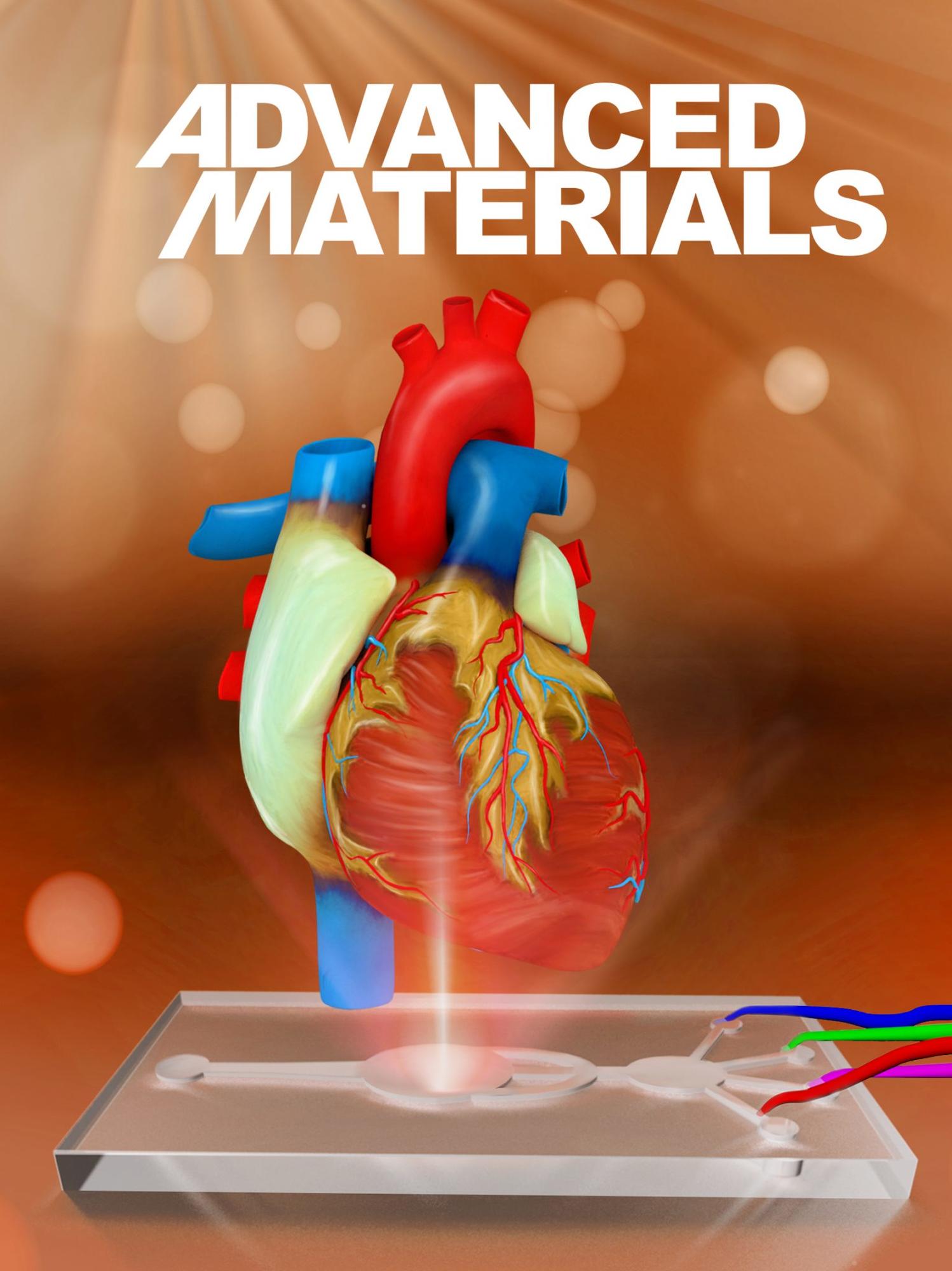
Miri AK, Nieto D, Iglesias L, Hosseinabadi HG, Maharjan S, Ruiz-Esparza GU, Khoshakhlagh P, Manbachi A, Dokmeci MR, Chen S, Shin SR, Zhang YS, and Khademhosseini A. 4/2018. “Microfluidics-Enabled Multi-Material Maskless Stereolithographic Bioprinting.” Advanced Materials. LinkAbstract
A stereolithography‐based bioprinting platform for multimaterial fabrication of heterogeneous hydrogel constructs is presented. Dynamic patterning by a digital micromirror device, synchronized by a moving stage and a microfluidic device containing four on/off pneumatic valves, is used to create 3D constructs. The novel microfluidic device is capable of fast switching between different (cell‐loaded) hydrogel bioinks, to achieve layer‐by‐layer multimaterial bioprinting. Compared to conventional stereolithography‐based bioprinters, the system provides the unique advantage of multimaterial fabrication capability at high spatial resolution. To demonstrate the multimaterial capacity of this system, a variety of hydrogel constructs are generated, including those based on poly(ethylene glycol) diacrylate (PEGDA) and gelatin methacryloyl (GelMA). The biocompatibility of this system is validated by introducing cell‐laden GelMA into the microfluidic device and fabricating cellularized constructs. A pattern of a PEGDA frame and three different concentrations of GelMA, loaded with vascular endothelial growth factor, are further assessed for its neovascularization potential in a rat model. The proposed system provides a robust platform for bioprinting of high‐fidelity multimaterial microstructures on demand for applications in tissue engineering, regenerative medicine, and biosensing, which are otherwise not readily achievable at high speed with conventional stereolithographic biofabrication platforms.
miri_et_al_advanced_materials.pdf
2017
Engineering a highly elastic human protein-based sealant for surgical applications.
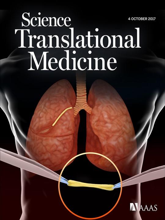
Annabi N, Assmann A, Vegh A, Ghasemi-Rad M, Bagherifard S, Cheng G, Ruiz-Esparza GU, Wang X, Noshadi I, Lassaletta A, Gangadharan S, and Khademhosseini A. 10/2017. “Engineering a highly elastic human protein-based sealant for surgical applications.” Science Translational Medicine, 9, 410. LinkAbstract
Surgical sealants have been used for sealing or reconnecting ruptured tissues but often have low adhesion, inappropriate mechanical strength, cytotoxicity concerns, and poor performance in biological environments. To address these challenges, we engineered a biocompatible and highly elastic hydrogel sealant with tunable adhesion properties by photocrosslinking the recombinant human protein tropoelastin. The subcutaneous implantation of the methacryloyl-substituted tropoelastin (MeTro) sealant in rodents demonstrated low toxicity and controlled degradation. All animals survived surgical procedures with adequate blood circulation by using MeTro in an incisional model of artery sealing in rats, and animals showed normal breathing and lung function in a model of surgically induced rat lung leakage. In vivo experiments in a porcine model demonstrated complete sealing of severely leaking lung tissue in the absence of sutures or staples, with no clinical or sonographic signs of pneumothorax during 14 days of follow-up. The engineered MeTro sealant has high potential for clinical applications because of superior adhesion and mechanical properties compared to commercially available sealants, as well as opportunity for further optimization of the degradation rate to fit desired surgical applications on different tissues.
annabi_et_al_science_translational_medicine.pdf

Pages